Article
In her master’s thesis, an AGH University alumnus, Dominika Ciupek, MSc Eng., described an innovative application of MRI diffusion imaging which combines tractography with the research on the nervous tissue microstructure. Conducted based on the presented methodology, the analysis provides new information on the process of brain maturation and ageing. The research was recognised in two competitions – “Diamenty AGH” and “Młodzi Innowacyjni”.
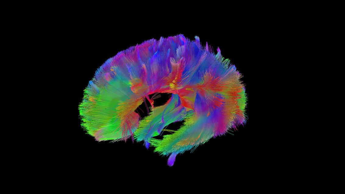
Tractography is one of the computational techniques taking advantage of diffusion MRI (Magnetic Resonance Imaging) which has been developing since the 90. It allows for mapping the properties of white matter pathways by providing spatial images with colour-coded paths. While observing such a tangle may be appealing to the eye, this is not what it is about. Such visualisations are used by doctors in diagnosing brain tumours and planning surgical procedures, however diffusion MRI is also a useful tool for neuroscientists studying connections between different areas of the brain and the functioning of the nervous system.
To create tractography images, the signal of diffusion-weighted MRI (DW-MRI) is registered by a scanner. The movement of water particles in the organism may take place in an isotropic or anisotropic way, when diffusing molecules meet limitations in the tissue. After transforming the signal with the use of a model representing the multidimensional structure of nervous tissue, the information on the character of diffusion may be obtained for each voxel. A commonly used representation of the signal is Diffusion Tensor Imaging (DTI), which measures the diffusivity along three perpendicular directions. It allows for establishing the main direction of the particles’ movement and describing the entire diffusion process with quantitative measures, among others. Some of them are Mean Diffusivity (MD) and Fractional Anisotropy (FA) which provide information on tissue microstructure, which allows to detect changes resulting from neurodegenerative diseases or natural aging processes.
How is a tractography image made?
Molecules diffuse with more freely along the nervous fibres than across them. A tractography image is created as a result of connecting neighbouring voxels of similar anisotropy, very likely to be from the same nervous fibres, with some help of the right mathematical and computer techniques. A diffusion tensor generalised to three directions is however only a mathematical object which does not truly represent the actual structure of nervous tissue (in reality, to determine a diffusion tensor, one has to make measurements at least in six independent directions). For brain areas where fibres are not parallel but, for example, intersect, the obtained images may be falsified. Biophysical models, which take into account certain assumptions about the structure of nervous tissue, perform better in this regard.
“Biophysical models take into account the existence of the so-called compartments,” explained Dr Tomasz Pięciak from Escuela Técnica Superior de Ingenieros de Telecomunicación at the University of Valladolid (Spain), former employee of the Faculty of Electrical Engineering, Automatics, Computer Science, and Biomedical Engineering at the AGH University, who is specialised in mathematical methods and computer algorhythm used for representing and processing data obtained in diffusion MRI. “In a certain compartment directional diffusion may be observed with free diffusion in the other, like between axons. We have a possibility of separating constituents of this signal. Still, the application of biophysical models requires more complicated and time-consuming methods of data acquisition, which exceeds the capacity of hospital diagnostics.”
Combination of tractography with microstructural research
What would elongate the queue of patients waiting for their results in the healthcare system is not an obstacle in the world of science. In her thesis entitled “Mapping the properties of white matter pathways of the ageing brain using the diffusion magnetic resonance imaging” developed at the Faculty of Electrical Engineering, Automatics, Computer Science, and Biomedical Engineering under the supervision of Dr Tomasz Pięciak and Dr Jaromir Przybyło, the AGH University graduate, Dominika Ciupek, described the use of an innovative method combining tractography with quantitative research to evaluate the microstructural changes occurring with age along four main pathways in the white matter: arcuate fasciculus, inferior longitudinal fasciculus, uncinate fasciculus, and corticospinal tract. The thesis was recognised in 2023 in competitions “Diamenty AGH” and “Młodzi Innowacyjni organised by the Łukasiewicz Research Network – Industrial Research Institute for Automation and Measurements PIAP, receiving the first prize in both of them.
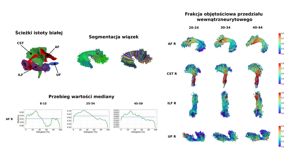
For the analysis, the thesis’ author used a base of MR scans from 62 heathy people in various age. By using a tractography algorhythm based on fibre orientation distribution (FOD), Ciupek established the fibre orientation in brain areas of her interest. Subsequently, on the basis of DTI, NODDI models (Neurite Orientation and Dispersion Density Imaging), and SMT (Spherical Mean Technique), the author determined structural measures along certain pathways. Due to high demand of computational power resulting from the biophysical models used in the research, the graduate took advantage of Prometheus’ resources, a supercomputer made available by the Academic Computer Centre CYFRONET.
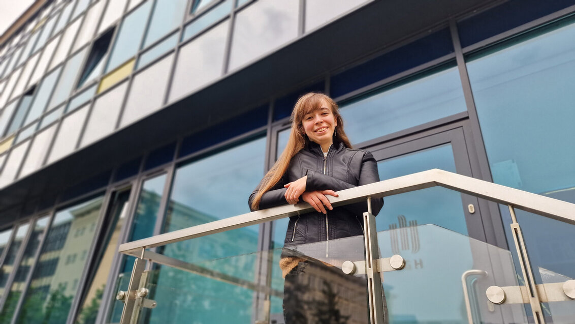
Dominika Ciupek, MSc Eng., photo by Marianna Cielecka
Connections in the brain mature and age equally
The conducted analysis allowed to make interesting conclusions: “The values of singular microstructural measures along certain fibre bundles change at an even pace throughout life. We cannot talk here about a quicker maturation or ageing of a front part of a bundle than of the back part. It is also not the case that measures only constantly grow or only decrease from the birth until the old age, instead they take the form of a parabola – once they reach their maximal value, it can be said that changes related to maturing stop and, after surpassing the extreme, the degeneration of a given bundle starts, i.e. the aging process. We can say that it occurs between the age of 25 and 35,” claims Dominika Ciupek, MSc Eng.
Dr Tomasz Pięciak emphasises that the methodology delineated in the thesis is innovative in comparison to other similar research aimed at capturing certain changes occurring in the brain. “We often learn from the media that scientists have studied how something, such as using social media or being around noise, affects the brain. What does it mean in practice? Most often, these researchers determine quantitative parameters on brain images and compare them with those obtained from a reference group. In this case, these measures were compared not within the area of interest, but along specific fibres designated with the use of tractography. Showing how they change over the course of life is the most important part of this thesis.”
In the quantitative assessment, the course of the median remains largely the same from one age group to the next, with only the range of values changing, figure by Dominika Ciupek
Federated learning as a new research direction
The AGH University alumnus continues to pursue her scientific interests related to microstructural measures within her doctoral research carried out in the international research institute Sano – Centre for Computational Personalized Medicine under the supervision of Dr Tomasz Pięciak and Professor Maciej Malawski from the Faculty of Computer Science at the AGH University.
In her research, Dominika Ciupek is focused on federated learning which makes it possible to improve the performance of AI used for various tasks in the field of MRI. One of the obstacles to the use of machine learning in this field is the fact that, due to the need to protect sensitive medical data, artificial intelligence models are trained on limited data resources, which cannot be shared by individual entities owing to current regulations. In the case of MRI, where scans taken from the same patient may vary depending on the manufacturer or equipment configuration, the difficulty is the replicability of the results produced by artificial intelligence on a wider scale.
“Federated learning allows each institution to teach their model separately with only the parameters being transferred and aggregated to get a better global model,” clarifies Dominika Ciupek, MSc Eng.
Previous achievements of the AGH University graduate
Awards for the thesis “Mapping the properties of white matter pathways of the ageing brain using the diffusion magnetic resonance imaging” are not the sole accomplishments of Dominika Ciupek, MSc Eng. In 2021, her engineering thesis prepared under the supervision of Dr Tomasz Pięciak, also on the application of biophysical models in the context of MRI, took the second place in the competition “Młodzi Innowacyjni” and in the Siemens Award competition in automatics and robotics. Ciupek was also recognised by the Ministry of Science and Higher Education and was awarded a grant for the continuation of conducted research as part of the “Best for the Best! 4.0” programme. Her thesis also received a distinction at the 30th Conference of Student Research Clubs “Człowiek i jego środowisko” in Kielce. Still prior to defending the thesis, a result of one and a half year’s cooperation with researchers from Italy and Iran, it was accepted to the prestigious ISMRM & SMRT Annual Meeting & Exhibition in Vancouver, Canada.
 Advance your career with a non-degree postgraduate programme at AGH University
  AGH University student to head the Student Council under the President of POLSA
  The first Krakow Solar Boat Challenge comes to a close
  AGH Solar Boat finishes 4th in the AI class at prestigious competition in Monaco
  AGH University International Faces – Episode #10: Yuka Shimizu
  AGH University with an impressive result in the evaluation of its scientific activity: seven disciplines with category A+
  Reaxys – a new resource in the Main Library subscription