Skip to content Skip to footer
News

Implants at the push of a button

A colourful illustrative image – a 3D image of a human skull.

Implants at the push of a button

Artificial intelligence will design cranial implants not only equally good as humans, but significantly faster – envisions Dr Eng. Marek Wodziński from the Faculty of Electrical Engineering, Automatics, Computer Science, and Biomedical Engineering. Tens of thousands of people can benefit from this project.

To fill a defect, all you have to do is design an implant that can replace the damaged bone. Then, using a material approved for medical use, manufacture its physical form and finally perform the replacement surgery. In the majority of cases, medicine sees no obstacles in successfully carrying out such surgeries – science gave us all the tools we need to help people with cranial defects.

However, in 2019, people in need of a cranial implant amounted to about 5 million patients around the world who, despite medical recommendations, had not undergone a neurosurgical procedure that would allow them to recover completely. Similarly, in Poland, many people who had been deprived of an intact mechanical barrier of the brain as a result of surgical intervention due to injury or cancer are forced to wait for an implant for months. During the wait, places and situations that for most of us are merely everyday nuisances can pose fatal threats to them. Moreover, such patients also struggle with social exclusion or reluctance to socialise because they feel different due to deformations. This long waiting time for a surgery has two main reasons – the process of manufacturing personalised cranial implants is currently extremely time-consuming and the number of neurosurgeons is insufficient.

Swiftness requires innovation

Implants are designed by bioengineers who are qualified to work with specialised software. Modelling an implant optimally adjusted to the defect in question requires them to individually analyse the medical data of a patient – this can take from several to several dozen hours. To make the design a reality, the project is usually sent to another company that produces the implant using, for instance, 3D printers; and then to yet another company that will sterilise it (if this procedure is not performed in a hospital). The entire process of approving the use of the implant can be considerably prolonged and take weeks or even months.

The AGH UST scientist, Dr Eng. Marek Wodziński, noticed the problem and decided to have a go at solving it. The National Centre for Research and Development, within the LIDER competition, rewarded his research project titled: Innowacyjny system do projektowania i weryfikacji spersonalizowanych implantów czaszkowych oparty o sztuczną inteligencję i mieszaną rzeczywistość [Innovative system for designing and verifying personalised cranial implants based on artificial intelligence and mixed reality]. The young scientist received PLN 1,500,000 to implement the project.

The innovation is that the system will automatically design a personalised implant for a given patient. Immediately in the operating theatre, a neurosurgeon will receive a tailor-made model ready to be printed by pushing a single button’, the project leader says about his goals.

Dr Marek Wodziński can already boast about a success related to a research activity in the medical industry – last year, he received the first prize in the ABB competition for his doctoral dissertation titled: Medical image registration methods focused on the problem of missing data, in which he proposed a series of algorithms aimed at increasing the quality of medical imaging and as direct support for medical procedures. Furthermore, for his research activity, he was awarded the START scholarship, granted by the Foundation for Polish Science, and a ministerial scholarship for exceptional young scientists.

Deeper insight brings a new quality
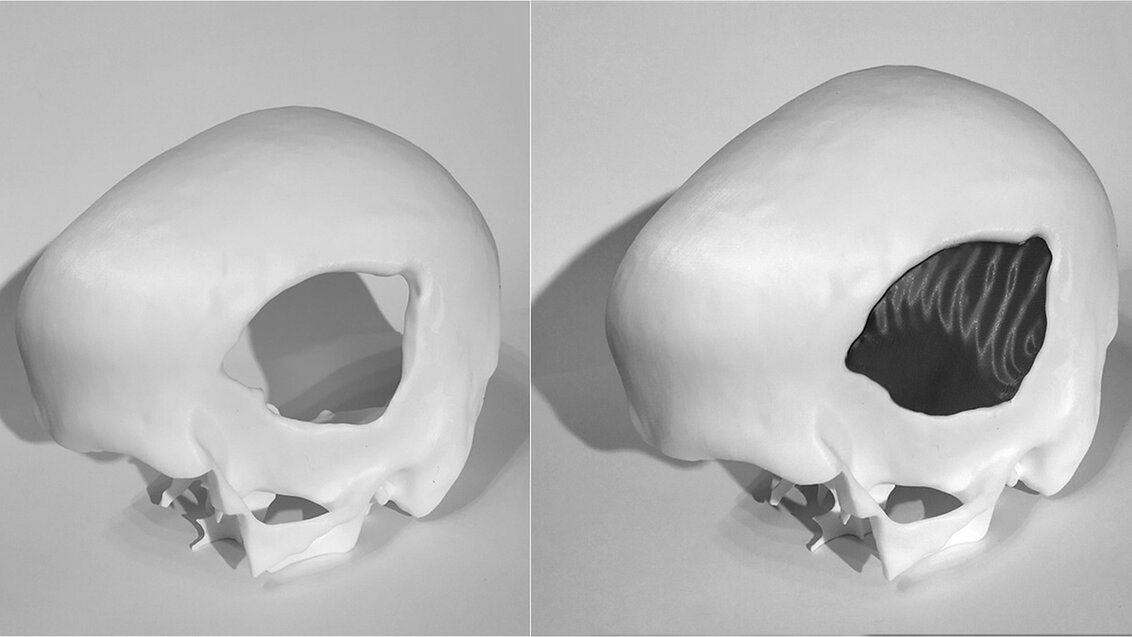
We’ve already designed algorithms that can successfully fill a cranial defect. The problem is that very often the cranial defect itself is not compatible with the implant, as the former can have a shape that physically won’t fit in the cranial cavity’, Dr Marek Wodziński says about the difficulties related to programming this innovative method. ‘Sometimes, as a result of traffic accidents, the skull is first damaged and then broken, which renders the situation more dangerous on the inside than on the outside – and we can’t put something in from the inside of the skull. Therefore, the replacement procedure requires further modifications to the defect model. Additionally, implants are made of different materials. So, depending on the material favoured by a given neurosurgeon, the implant will yield different mechanical and different biological responses – this must also be taken into account during the design stage. Some implants will have to be thicker, others thinner; they are also fitted in various ways. We must also consider the fact that immediately after the procedure the brain can be deformed and we may have to deal with swelling. Implants should not cause an increase in intracranial pressure; therefore, they should always be a bit thinner than the original cranium. There’s still a long way to go from the defect to the implant’, explains the scientist.

Previous attempts at creating algorithms to design implants by other research teams have yielded moderate results. These algorithms can deal quite well with designing implants for defects similar to those they had analysed before, but cannot transpose their expertise to new cases with which they had not dealt before.

The project will definitely have to account for increasing the ability to generalise these proposed algorithms to include cases that had not been shown to the software during training’, says the project leader.

Two illustrative images: the one on the left shows a human skull with a large defect; the right one shows the same skull fragment, but the defect has been filled with an implant.

Tackling problems in advance

To arrive at a breakthrough, the scientist wants to use artificial intelligence, especially deep learning algorithms based on artifical neural networks. Deep learning is a way of data processing that started to be increasingly popular about a decade ago and still finds new applications in places where large collections of data need to be analysed. Although its origins can be traced back to drawing inspirations from the structure of the human brain, nowadays, as the scientist himself claims, the group of algorithms designated as such does not exclusively involve a simulation of biological foundations of thought processes.

First neural networks have been inspired by nature, by the way the human brain works. Now, however, it all took a slight turn, as this group of algorithms models complex non-linear functions. They still draw inspiration from nature and the process of transmitting signals within the human brain, but it became somewhat simplified, so I wouldn’t look for direct analogies’, says Dr Marek Wodziński.

Deep learning fuels conversation bots and autonomous vehicles, but it can also teach a program to recognise cancer cells. In this project, the aim of the algorithm focuses on a comprehensive design of a personalised implant. Data that are supposed to be used to create the design may come from images taken during a CT scan or an MRI. The latter method of acquiring information about the defect is especially useful in patients with tumours, as magnetic resonance is a standard medical procedure in cancer diagnostics, so obtaining an image would not overcomplicate the procedure. Moreover, MRI is a non-invasive method.

In relation to implant design, the fundamental advantage of deep learning algorithms is that they can quickly produce the desired responses. The project leader sees an opportunity not so much as to reduce the wait, but to eliminate it completely – this would be possible if designing, printing, and sterilising the implant took a sufficiently short time, so that surgeons would not have to wake patients from anaesthesia and replace the defect during the same surgery in which the bone fragment had been removed. Instead of two surgeries, we would have only one, which significantly reduces the risk of complications for the patient and reduces the time to replace the bone defect. Therefore, this solution can prove especially tempting for investors from the United States, where neurosurgeons make about 600,000 dollars. Therefore, every hour of their time is worth a lot not only in the eyes of people waiting for a surgery, but also in the eyes of the surgeons’ employers. However, savings would only become a reality if the implant had been made directly during surgery.

Obviously, we could propose other algorithms, but other numerical solutions are often quite time-consuming. The design of an implant can take even a few hours. Deep learning allows us to transfer the entire time-consuming calculation stage to the training process – a neural network is first taught to perform a given task, e.g. how to reconstruct a cranial defect and then how to design a specific implant. Thanks to that, using it as a tool is a matter of seconds. In this way, we can introduce innovation: not only will the system design really good implants, but it will also do it efficiently, becoming useful to doctors or directly in the operating theatre'.

Reward in the form of stimulation

The scientist wants to use a subgroup of data processing methods in the field of deep learning that includes methods rarely applied to problems regarding medical imaging, that is, reinforcement learning. These methods acquire data because the ‘environment’ reacts to their actions. Bots are created in this way, which are able to defeat humans in various games, including chess. This was also the learning pattern used to teach artificial neuron cultures to play Pong, a simple game in which you have to hit a ball back and forth between two paddles so that it does not escape the game field. What was the ‘carrot’ for the neurons? The researchers determined that predictability will be the reward for each system, which is why when the movements of the algorithm, following the rules, allowed it to score a point, the researchers stimulated the network exactly in the same way. If it did something out of line, the tissue stimulation was random. Eventually, neurons started to react as if they ‘wanted’ to win, so the learning process seemed to be a success.

In the case of Dr Marek Wodziński’s research, the ‘environment’ of the algorithm should therefore react one way when it proposes a correct implant and another way if the results of the calculations are not satisfactory enough for a replacement procedure. This will be the path to acquire data on the models that could be attached to the cranium. If the algorithm processes enough cases, it will be able to adjust an implant to any defect.

Mending, fitting, and a different reality

When a doctor ‘pushes a button’, they will generate an implant with matching parameters; but, before printing, the system will give the user the opportunity to easily modify the designed solution with a user-friendly interface. All this to make it easier for doctors and, at the same time, to expedite the entire process and increase the efficiency of surgeries. Every surgeon can have slightly different preferences regarding the shape and size of the implant, depending on the technique of surgery: the system cannot foresee it, as there may be countless implants that will fulfil their function, and each surgeon will choose differently.

If a doctor wants to modify something, for example, they want to have a wider or narrower implant with a different bone dialation, the system will allow them to manually control the design process. However, they will not have to use any specialised software. Instead, they will just have to move a few sliders with which they’ll be able to adjust specific parameters’.

Dr Marek Wodziński highlights that he introduced other amendments to the project, which are to boost the commercial attractiveness of the solution. These boosters include the use of mixed reality in the process of filling a defect. The doctor will have the option of wearing special goggles that will still allow them to see physical objects in the room, but also a hologram of the designed implant. This will allow them to ‘fit’ it to the cranium even before the implant is physically produced. This means convenience and savings – the necessity of possible amendments will be immediately spotted and introduced, and the costly materials used to print out the implants will not go to waste until the operator is certain that the implant has exactly the shape it should.

Enough for science, not enough for implementation

Where will the LIDER competition money go? Most of the funds will be allocated to purchasing equipment and materials necessary to develop and test the efficiency of the new solution.

The calculation infrastructure is definitely a considerable cost. We have computers at the AGH UST, Prometheus and Athena, for instance, but they are devices that – at least according to our research team – are used to make final calculations, specific comparative experiments, and trainings of final neural network models. Alas, it is not fitting equipment to prototype solutions – the interfaces of these computers are simply not made to do this. Therefore, we have to buy the necessary equipment. Other costs include compensation, as the project shall employ six people in total, which is why even small salaries amount to a significant portion of the money. Additional costs include the purchase of mixed reality sets and other minor expenses, such as managing a project website, promoting the solution, participating in scientific conferences, and working on open-access publications'.

Within the project, the team will use the equipment available at the AGH UST to print experimental implants which will then be presented to neurosurgeons from various countries for assessment of their efficiency and usefulness in practice. At this stage, the implants will not yet be used in patients – although the scientists plan to also use medically certified materials to print the bone implants. However, to place the implants designed in patients, the team would need to get the software certified. As Dr Marek Wodziński says, this step significantly transcends the budget of this project – we would really need additional funding from the AGH UST or an external investor. The project leader adds with a pinch of optimism that obtaining an approval for the software is a much shorter process than obtaining an approval for printers or biomedical materials, which could take years. Using this equipment simplifies things a lot – if we can only create a functional prototype and find a funding source, obtaining an approval should be fairly quick. This piece of news should uplift not only people who wait for cranial reconstruction, but also us all – because science is about to push back another frontier.

Stopka