News
Illustrative image. Photo: Dreamstime
Artificial intelligence can remove artifacts and noise from images caused by interference in signal acquisition by comparing them with a reference image. However, it is the manner in which the latter is generated that largely defines whether other fragments that could be significant in diagnosing patients are removed. A method described in a diploma thesis by an AGH UST student deals with the problem much better than the solutions currently in use.
Diffusion magnetic resonance imaging is one of the diagnostic techniques based on the phenomenon of nuclear magnetic resonance, which is particularly useful in brain scans. The technique relies on the diffusion of water particles, which can be isotropic and anisotropic, where the movement of molecules is constrained by natural barriers. For instance, particles diffuse with much greater freedom along the nerve fibre axes than across them. Diffusion imaging for each voxel, that is, the smallest unit in three-dimensional space, facilates the characterisation of this process with quantitative measures. Therefore, unlike traditional magnetic resonance imaging, the images obtained in this way are secondary to the numerical parameters determined during the scan.
Diffusion imaging can be a great tool for detecting cancer and neurodegenerative diseases or to track the natural changes that occur in the brain during the ageing process. Regrettably, the relatively long time required to perform the scan limits the usefulness of this technique in clinical practice. The signals collected by the scanner can be corrupted by noise due to, for example, changes in a patient’s body temperature or heart rate, or interference caused by thermal noise. As a result, to obtain images free from deformation, the acquisition should be repeated multiple times.
Reference or pseudo-reference images?
Current scientific investigations that aim to obtain high-quality images with a reduced number of necessary repetitions focus on the use of artificial intelligence methods to denoise data. Convolutional neural networks can learn to remove distortions by comparing images corrupted by noise with reference images or, more precisely, pseudo-reference images. This is important because a raw signal registered by the scanner is not useful in any way to the person interpreting the results without proper processing of data using mathematical tools and computer algorithms. To do this, we can use models that represent the multidimensional structure of nervous tissue.
‘Choosing the right mathematical model is crucial and facilitates the proper quantitative presentation of the diffusion process within the brain area. Standard models can be successfully applied in clinical practice; however, they might overlook significant diagnostic data. On the other hand, using more advanced models prolongs the time a patient has to spend in the scanner; and in some cases, it requires making the additional choice of a dedicated data acquisition protocol’, explains Dr Eng. Tomasz Pięciak from the Faculty of Electrical Engineering, Automatics, Computer Science, and Biomedical Engineering and the Escuela Técnica Superior de Ingenieros de Telecomunicación at the University of Valladolid (Spain), who specialises in mathematical methods and computer algorithms used to present and process data obtained in the process of diffusion magnetic resonance imaging.
Diffusion tensor vs. spherical harmonics
The ability of artificial intelligence to effectively denoise images will not achieve much in clinical practice if an inappropriate set of data is used to train it. In such a situation, relevant diagnostic data might be removed from even good-quality images. A step forward in relation to the currently available methods has been proposed by Julia Machnio, Eng. in her diploma thesis written at the Faculty of Electrical Engineering, Automatics, Computer Science, and Biomedical Engineering under the supervision of Dr Tomasz Pięciak.
‘The thesis manages to combine fire with water, that is, using complex models for acquisition times that are close to those commonly applied in clinical practice, while simultaneously preserving the compatibility of procedures’, the supervisor comments.
The starting point for Julia Machio’s considerations came from a method that had already been proposed in the literature, namely DeepDTI. The authors of the paper generated pseudo-reference data using DTI (diffusion tensor imaging). The neural network they trained learned to denoise images in a relatively short time, which allowed them to obtain visually high-quality images.
‘The problem is that tensor modelling is limited by the direction of diffusion. To put it overly simply, if a cancer does not fit the model, it will not be correctly classified. This was the most serious drawback of the aforementioned work, on which we wanted to focus on’, Julia Machnio explains.
The DeepSH method she proposed in her work to generate pseudo-reference data uses an alternative method of spherical harmonics.
‘We used the same data, but we modelled it using a different function that also takes into account the diffusion signal with a more complex architecture’, describes the author of the thesis.
A method more reliable than others
The investigation carried out by Julia Machnio provided proof for the high efficiency of the method to denoise images, while simultaneously preserving data in the processed signals, which might carry significant diagnostic information.
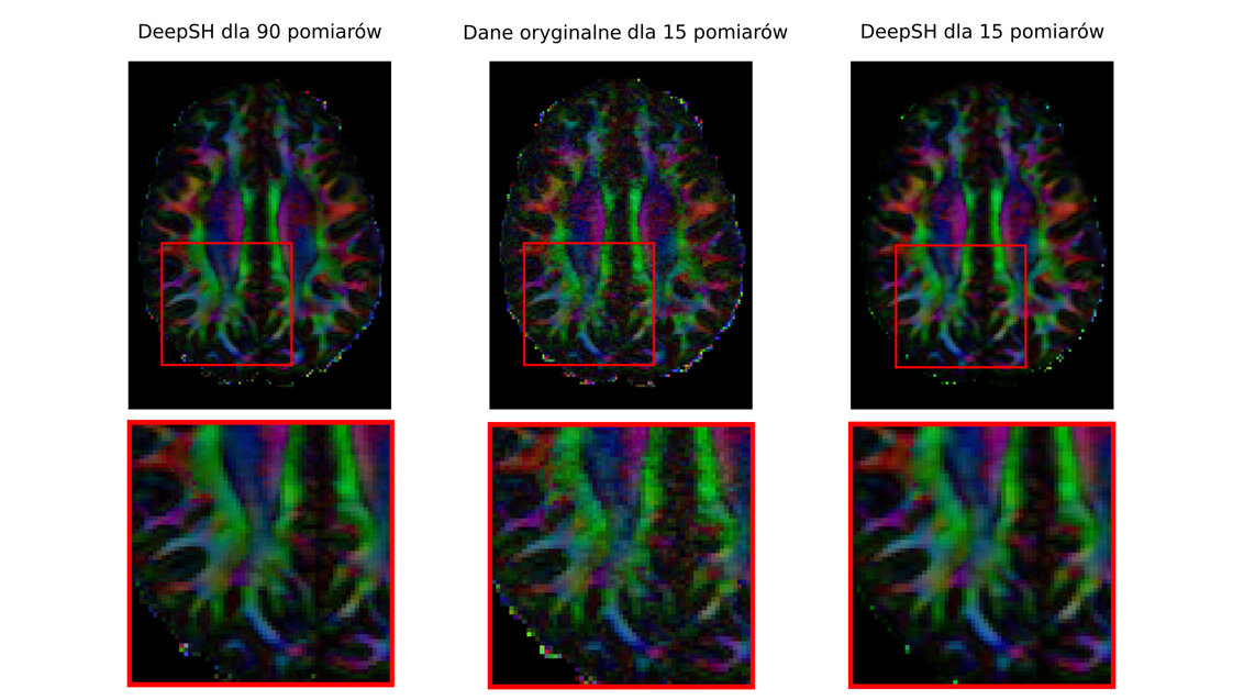
‘The differences between the image obtained for the spherical harmonics and the real image were less than 10%. Whereas for the tensor, in some cases, the differences amounted even to 20–30 per cent. When such a significant portion of the signal differs from the image, which has not been processed in any way, we should bear in mind that what had been removed was not only the noise, but also a medically vital signal’, reports the AGH UST student.
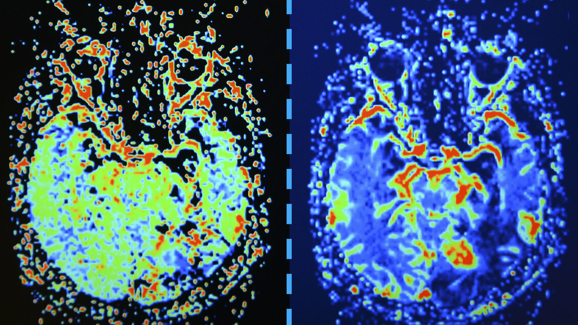
A pseudo-reference image and data before and after denoising using the DeepSH method
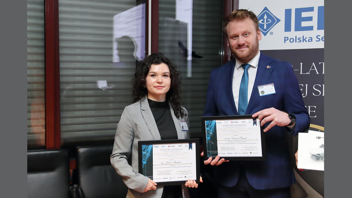
Julia Machnio, Eng. accepting the award in Warsaw during the 50-year anniversary of the IEEE Poland Section. Photo: IEEE Poland
 Begin your journey at AGH University Doctoral School
  Advance your career with a non-degree postgraduate programme at AGH University
  Opus LAP grant for a Polish–Czech project
  AGH University among top institutions in renowned Perspektywy University Ranking
  Universities join forces to launch Inter-University Space Ecosystem Center
  Protein research to inform the search for life in space
  AGH University visits Peru. Three new agreements with PUCP